What are the world’s top 5 magnetic resonance clinical research of 3T MRIs
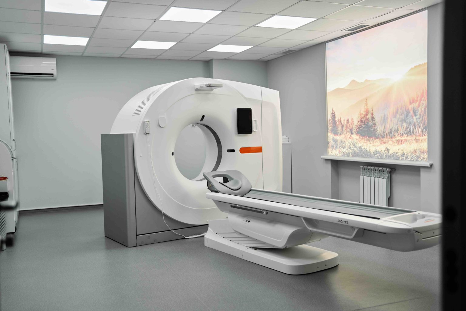
MRI is a type of diagnostic test that can create detailed images of nearly every structure and organ inside the body. MRI uses magnets and radio waves to produce images on a computer. MRI does not use ionizing radiation. Images produced by an MRI scan can show organs, bones, muscles and blood vessels.
In July 2009, the Human Connectome Project (HCP), funded by the National Institutes of Health (NIH), was officially launched. As the highest-level research project in the field of neurology, it aims to use different brain imaging techniques (such as functional magnetic resonance imaging, diffusion magnetic resonance imaging, supplemented by EEG, MEG, etc.) to draw “network diagrams” of different living human brain functions and structures to clarify the anatomy and functional connectivity in the healthy human brain, which will not only help to understand the mysteries of thoughts, feelings and behaviors in the brain, but also generate a large amount of data that will help study brain diseases.

To discover SECRETS of How to get most Cost-effective Medical Equipment.
Why Perlove Medical 007 Quality 100% Competetion: ***Siemens, GE, Philips, Medtronic, Shimadzu, Fuji Film etc***
Why more Medical Distributors choose Medical 007 over other VENDORS in the world?
Wanna exact details, just click:
PERLOVE 007: https://linktr.ee/medical86
SGS Audit Video: https://chdz.co/PERLOVE-V
ATTN: Jason Xie
Mobile Wechat & WhatsApp: 008613958401559
#MedicalEquipment #Healthcare #XRAY #CARM #DR #Fluoroscopy #angiography #Medicalinnovation #Siemens, #GE, #Philips #Medtronic #Shimadzu, #GMM #FUJIFILM
#MRI Scanners
#CT Scanners
#Ultrasound
#Radiotherapy and #Radiosurgery
#Nuclear and #Molecular Imaging
Molecular Imaging #Facility, #Operations and Maintenance
X-Ray
Radiology and #Interventional
Injectors, Dispensers and Accessories
IIS/IT and Printers
The entire project is divided into two teams, one of which is the MGH-USC team led by Massachusetts General Hospital and the University of California, Los Angeles. In order to better collect data, they creatively developed an ultra-high gradient field magnetic resonance: Connectome 3T, which is a 3.0T magnetic resonance with a 60cm aperture, a maximum gradient strength of 300mT/m and a maximum conversion rate of 200T/m/s, a single FOV single scan 64-channel RF receiving platform, and a 64-channel head coil.

The reason why 7T was not selected is that: 1) The contrast of diffusion magnetic resonance imaging (dMRI) depends on the random displacement of water molecules when the gradient field is applied, which is independent of the main magnetic field strength; 2) Although the image signal-to-noise ratio is highly correlated with the field strength, the shorter T2 relaxation of 7T magnetic resonance partially offsets the high SNR brought by the high field strength; 3) Under the same parameters and subjects, the SAR value of 3T is less than 20% of that of 7T; in addition, compared with 7T, the interaction between the magnetic field and the gradient field of 3T is easier to handle, which also helps to reduce vibration and noise problems.
At present, there are only four Connectome 3T in the world, located in Massachusetts General Hospital in the United States, the Brain Imaging Research Center of Cardiff University in the United Kingdom, the Max Planck Institute for Human Cognitive and Brain Sciences in Germany, and the Zhangjiang International Brain Imaging Center of Fudan University in China.
Distribution of four Connectome 3T MRIs in the world (from the Internet)
Although the Connectome 3T MRI equipped with 300mT/m@200T/m/s ultra-high performance gradient cannot be used directly in clinical practice, as a “by-product” of HCP gradient development, the gradient performance of commercial MRI has been continuously enhanced in the past decade, providing a strong hardware foundation for high b-value diffusion, brain functional imaging, multi-layer simultaneous imaging and micron imaging.
For medical institutions, the ideal clinical research MRI should first serve the clinic and be fully capable of taking into account all kinds of scientific research. In order to facilitate hospitals to understand the latest MRI products, the best clinical research 3.0T MRIs are listed in the order of release time (some brands have two, which are comparable).
1. Siemens MAGNETOM Prisma & Vida
Product Introduction
1. MAGNETOM Prisma
In 2012, Siemens launched the 60cm aperture ultra-high gradient performance MRI: MAGNETOM Prisma. Although it has been released for 10 years, it is still one of the 3.0T MRIs with the highest hardware performance to this day.
Prisma is derived from its 7T platform, with a uniform magnet equivalent to 7T, the same ultra-high performance gradient of 80mT/m@200T/m/s as 7T; the same multi-source RF parallel emission TrueShape platform as 7T, and the same single FOV single scan 64-channel RF receiving platform as 7T.
As an imaging device recognized in the field of international brain science research, Prisma can be equipped with a 64-channel neurofunctional coil for brain science research, which makes the signal-to-noise ratio and resolution of the image reach the extreme of 3T magnetic resonance, so that its spatial resolution can reach 0.8*0.8*0.8mm, the time resolution can reach 0.5s, and the diffusion gradient can reach up to 514, which can meet the research of cortical substructure, cognitive science research and children’s brain development research.
2. MAGNETOM Vida
In 2017, Siemens launched the first life-sensing magnetic resonance in ECR: Vida (“Big Vida”), and realized localization in 2019. The hardware is equipped with a 70cm large aperture, 60mT/m@200T/m/s intelligent precision gradient system, the latest integrated coil technology: integrated life matrix coil system, and realizes a maximum of 204 receiving channels and a single FOV single scan with a maximum of 64 channels.
The biggest highlight of Vida is that it is equipped with the BioMatrix System, which integrates the latest physical technology, intelligent sensor technology and intelligent technology to achieve “MRI with patients”, so that patients can lie on the scanning bed, and the MRI can truly feel the patient’s anatomy, breathing, magnetic field and other body information, which not only greatly improves the comfort of patient scanning, but also ensures the imaging quality of moving organs such as the heart.
In addition, Vida is equipped with the Turbo Suite, which integrates magnetic resonance acceleration technology (parallel acquisition GRAPPA + simultaneous multi-layer acquisition SMS + compressed sensing Compressed Sensing), clinical scanning strategy, BioMatrix life perception and intelligent technology to meet the needs of different clinical and scientific research acceleration.
Siemens Prisma and Vida (from the Internet)
Core parameters
Innovative technology
In recent years, Siemens has also launched two technologies that are more conducive to high-throughput inspection and high-quality images:
1) Deep Resolve, a deep learning image reconstruction algorithm launched in 2022, can significantly shorten the scanning time and reduce noise at the same time, thereby improving image quality. For example, Deep Resolve can reduce the scanning time of brain MRI by 70% and increase the resolution by 1 time at the same time.
2) Contour Coils, ultra-flexible MRI coils also based on high-density lightweight coil technology.
As of now, the above two technologies have not been approved by the National Medical Products Administration (NMPA).
Siemens Contour Coils (from the Internet)
2. GE Healthcare SIGNA Premier
Product Introduction
In 2017, GE Healthcare launched a high-end MRI: SIGNA Premier, which is currently the only clinical research MRI that combines a large aperture of 70cm and an ultra-high performance gradient of 80mT/m@200T/m/s. As GE’s most advanced 3.0T MRI, it has five major advantages:
1) Ultra-uniform magnet: equipped with the industry’s highest third-order uniform field technology, it realizes dynamic correction and scanning of stable B0 fields, and increases the resolution and signal-to-noise ratio of whole-body diffusion imaging technology by more than 1.5 times.
2) SuperG Super Gradient: Equipped with the industry’s highest third-order eddy current self-calibration technology, it effectively overcomes the eddy current effect generated between the gradient switching process and the main magnetic field, and increases the utilization rate of the effective gradient from the traditional 66.7% to 86.7%.
3) 146 channels: It adopts a new 1:1:1 (1 acquisition channel corresponds to 1 analog-to-digital converter corresponds to 1 optical fiber) RF architecture, realizes a single-view 64+ full-body scan, and is equipped with the “Orchestra” symphony image reconstruction platform, which can achieve 81,000 frames/second high-definition reconstruction.
4) AIR platform: The same AIR technology platform as GE Architect.
5) Scientific research platform: The new QuantWorks holographic quantitative magnetic resonance platform realizes the quantification of both function and structure, bringing more refined and richer functional images and quantification. It should be pointed out that in terms of cardiac magnetic resonance, GE Healthcare and Arterys have launched 4D intelligent imaging technology: ViosWorks, which can present the heart from 7 dimensions: 3 spatial dimensions, 1 time dimension, and 3 speed dimensions. It can not only present the entire cardiovascular and cardiac structure, but also present the real-time status of the entire chest cavity in the form of video.
GE SIGNA Premier (from the Internet)
Core parameters
Innovative technology
In recent years, GE Healthcare’s most eye-catching innovation is the AIR platform, including AIR Coil, AIR Touch, AIR thunder, AIR X, and AIR Recon DL. It is a “package” solution that combines magnetic resonance signal acquisition, transmission, and reconstruction, representing the technical direction of GE magnetic resonance. For more information about the AIR platform, please refer to: What are the technical advantages of GE magnetic resonance AIR platform?
Among them, AIR Coil represents high-density lightweight coil technology, AIR Touch represents intelligent workflow, AIR thunder represents high-speed and high-quality acquisition, AIR X and AIR Recon DL are based on deep learning technology, AIR X realizes intelligent layer selection to ensure image consistency; AIR Recon DL achieves a significant reduction in scanning time while improving image quality.
So far, except for AIR X and AIR Recon DL, the others have been approved by NMPA.
GE AIR platform (from the Internet)
III. Philips Ingenia CX & Elition
Product introduction
1. Ingenia CX
Ingenia CX is a research-based MRI launched by Philips in 2017. It was put into use in the Department of Radiology of Peking Union Medical College Hospital in September of that year. As Philips’ first 60cm aperture research-based 3.0T MRI equipped with whole-body compressed sensing technology:
In terms of hardware, 1) it uses the Alpha intelligent gradient of 80mT/m@200T/m/s, with four intelligent modes: high resolution, high signal-to-noise ratio, stable and low consumption, silent and low heat. Intelligent combination of gradient field strength and switching rate to meet different clinical and scientific research needs; 2) it uses Multitransmit 4D multi-source RF transmission technology and dStream all-digital RF receiving technology to improve the image signal-to-noise ratio by 40%.
Functionally, 1) Micron imaging: Microscope technology combines high-performance intelligent gradients with high-definition digital microscopic coils to achieve micron-level fine imaging of anatomical structures; 2) Compressed sensing (Compressed SENSE), which can be applied to a variety of clinical and scientific research sequences throughout the body, and the image quality obtained is the same or better in 1/2-1/4 of the time of conventional scanning. 3) 3D-APT (amide proton transfer imaging), which uses high-resolution images to evaluate the protein expression of major diseases such as tumors, strokes, and geriatric diseases, providing important information for clinical diagnosis and treatment; 4D ASL technology can achieve drug-free DSA-like imaging, and can perform unilateral small field of view marking, providing a new method for the study of intracranial arterial blood supply areas.
2. Ingenia Elition
In 2018, Philips launched the top scientific research 70cm large-aperture magnetic resonance at ECR: Ingenia Elition, which will be domestically produced in October 2022.
Elition adopts the new Vega HP gradient design, using high-precision water jet cutting instead of traditional manual winding to make gradient coils, achieving a 220T/m/s gradient switching rate and 0 eddy current imaging, shortening TR and TE times. It provides up to 23% temporal resolution in functional magnetic resonance imaging (fMRI) studies, increases diffusion weighted imaging (DWI) scanning speed by 30%, and increases contrast resolution by an average of 70%. In addition, combined with compressed sensing, the scanning speed can be increased by 50%, or the spatial resolution can be increased by 60% within the same scanning time.
In addition to all the software and hardware technologies of Ingenia CX, Elition also innovatively introduced the Ambient Experience system, which features sound and light environment and noise reduction and mute technology, integrating architectural style, decorative patterns and enabling technologies (such as dynamic lighting, video projection and sound effects), which can make the patient’s medical environment more personalized and create a relaxing atmosphere.
Philips Ingenia CX and Elition (from the Internet)
Core parameters
Innovative technology
At the end of 2021, Philips launched a number of star technologies at RSNA:
1) SmartSpeed, an artificial intelligence reconstruction platform: Philips’ next-generation magnetic resonance acceleration technology for compressed sensing acquisition technology, based on the original adaptive intelligent compressed sensing algorithm (Adaptive-CS-Net), covers the entire process from acquisition, reconstruction, and post-processing.
2) Fully automatic workflow engine MR Workspace: With AI-assisted functions, it aims to simplify the entire process from image acquisition to diagnosis, speed up the turnaround time between patients, and improve the efficiency and productivity of radiology scheduling.
3) Multi-nuclear magnetic resonance imaging: Its latest high-end 3T magnetic resonance: MR 7700’s innovative technology has now been expanded to five other cell nuclei such as phosphorus 31 and sodium 23, which can improve clinical confidence through more metabolic and functional information, and make multi-nuclear imaging part of the daily magnetic resonance workflow.
So far, the above three technologies have not been approved by the National Medical Products Administration (NMPA).
Philips SmartSpeed achieves full process coverage (from the Internet)
IV. Canon Medical Vantage Centurian
Product Introduction
In 2019, Canon Medical launched the high-end MRI: Vantage Centurian, which is the industry’s first commercial clinical research MRI equipped with 100mT/m@200T/m/s. Both the software and hardware are the epitome of Canon MRI:
1) Double Hundred Gradients: With the Saturn X gradient system with high-voltage integrated gradient design, the ultra-high performance gradient of 100mT/m@200T/m/s not only realizes scientific research freedom, but also reduces the vibration amplitude of the gradient coil by 75% and the eddy current by 60% to improve image quality. In addition, the triple embedded water cooling (Direct Cooling) design improves the heat dissipation efficiency by 55%, ensuring that the gradient coil can work stably for a long time;
2) Pure source radio frequency: In addition to the use of multi-source radio frequency, it is also equipped with a pure source radio frequency ring, which can greatly absorb the electromagnetic wave interference caused by external noise mixing and gradient eddy current, eliminate signal source impurities from the source, improve the purity of the radio frequency field, and can increase the image signal-to-noise ratio by an average of more than 40%.
3) Super silent: Equipped with “Pianissimo Zen” hardware noise reduction technology, the gradient coil is placed in a vacuum cavity to block the sound from propagating through the air. Compared with conventional software and hardware noise reduction, combined with the integrated molding gradient “Pianissimo Zen”, the minimum superimposed scanning noise is only 2dB without extending the scanning time and reducing the image quality, and the maximum scanning noise can be reduced by 90%, improving the comfort and success rate of the examination;
4) High-definition cardiopulmonary imaging: Thanks to the unique pure source radio frequency technology, Centurian can improve the effective use rate of radio frequency signals, improve the signal ratio to ensure the signal-to-noise ratio and resolution. In addition to routine clinical applications, it also has a number of advanced clinical technologies, such as the first lung imaging in China to pass NMPA certification, which can non-invasively, radiation-free, and accurately evaluate lung parenchyma and functional changes; high-quality coronary imaging without contrast agent based on Sure ECG technology, which greatly improves the success rate of examination.
5) High-speed acquisition: Equipped with compressed sensing technology Compressed SPEEDER and artificial intelligence engine reconstruction algorithm AiCE, it can significantly shorten the examination time to achieve 60-second rapid imaging of the whole body while effectively separating image noise to obtain high signal-to-noise ratio images.
6) Other innovations: Equipped with “Sky Eye” technology, it can achieve accurate patient positioning and scanning, simplifying the magnetic resonance examination process; equipped with “The MR Theater” similar to Philips’ quiet environment experience system, combined with “Pianissimo Zen” silent technology, so that patients can watch movies while undergoing the examination to obtain a comfortable examination experience.
As of now, Vantage Centurian has not yet obtained approval from the National Medical Products Administration (NMPA).
Canon Centurian’s efficient “Sky Eye” scanning (from the Internet)
Core parameters
Innovative technology
In 2016, Canon acquired Toshiba Medical for US$5.7 billion and became its wholly-owned subsidiary, and changed its name to Canon Medical. Since then, it has continued to increase investment in the field of magnetic resonance to accelerate the growth of its magnetic resonance business. For example:
1) Acquired Olea medical, a French company specializing in CT and MR image post-processing solutions, and seamlessly integrated its innovative neurology, oncology, sports medicine, cardiology and women’s health and other rapid and accurate diagnostic solutions into its Vitrea post-processing workflow to enhance diagnostic confidence;
2) Acquired Quality Electrodynamics (QED, also a supplier of some magnetic resonance coils for GE and Siemens), a world-leading magnetic resonance radio frequency coil company, mastered all the technologies of high-density lightweight coils, and also launched its coil blanket Shape Coil;
3) Acquired Skope, a Swiss company specializing in high-quality magnetic resonance imaging, and is committed to combining sensor technology with MR signal processing and image reconstruction to bring more accurate and detailed magnetic resonance imaging. Among them, one of Skope’s technologies is similar to KINETICor (Siemens Life Perception Biomatrix supplier). Through the sensing camera, it can directly sense the dynamic changes of the magnetic field and the patient’s anatomical, respiratory and other physiological information to obtain better examination results.
Application of Olea post-processing technology in sports medicine (from the Internet)
V. United Imaging Medical uMR880 & 890
Product Introduction
In 2020, United Imaging Medical released the 60cm aperture clinical research 3.0T magnetic resonance uMR890, equipped with the industry’s highest commercial 120mT/m@200T/m/s ultra-high performance gradient; in 2021, United Imaging continued to release the high-end 3.0T magnetic resonance uMR880, which are currently the two most advanced magnetic resonances of United Imaging. Since the details of uMR890 cannot be determined (it is speculated that it has several more acquisition channels than uMR880, and the others are the same), the uMR880 is mainly introduced:
In terms of hardware, 1) This is a 65cm aperture MRI, and its magnet adopts horizontal fish scale welding and other processes to achieve excellent magnetic field uniformity, more uniform fat compression effect, smaller diffusion deformation, more accurate spectral imaging, and functional imaging. 2) With the support of 3.5MW high-power gradient amplifier, it achieves 80mT/m@200T/m/s high-performance full digital gradient, laying a solid foundation for advanced imaging of all parts of the body. Taking neuroimaging as an example, it can meet the standards in the HCP scanning protocol; 3) It is equipped with a high-density super-flexible coil (SuperFlex Coil) similar to GE Magic Carpet, and its unit density is increased by more than 50% compared with the previous generation.
In terms of functions, it is equipped with the biggest innovation in the field of magnetic resonance imaging in recent years: the uAIFI Technology platform, which realizes intelligent scanning, greatly improves imaging speed and signal-to-noise ratio, and gives patients a more comfortable examination experience, including: 1) ACS and DeepRecon, which can achieve hundred-second imaging of all parts of the body, saving an average of 80% of scanning time, and intelligently identify and remove noise while ensuring the details of image diagnosis, further improving the image signal-to-noise ratio; 2) uVision, EasyScan and EasySense, apply the Sky Eye technology to magnetic resonance scanning, and combine it with the perception drive technology similar to Siemens Life Perception Matrix, which can realize non-contact physiological movement perception, complete one-button intelligent scanning of multiple parts of the body, covering more than 80% of clinical scenarios; 3) Post Processing, based on artificial intelligence technology, simplifies some post-processing, realizes intelligent cropping, intelligent plaque analysis, and intelligent brain analysis.
United Imaging uMR880 (from the Internet)
Core parameters
Innovative technology
For United Imaging, magnetic resonance imaging is its trump card business, and it has always been highly valued and invested heavily. In recent years, we have launched China’s first 75cm ultra-large aperture 3.0T MRI: uMR OMEGA, China’s first 5.0T MRI: uMR Jupiter, China’s first ultra-high field animal MRI: uMR 9.4T, and the innovative uAIFI Technology platform.
United Imaging uAIFI Technology Platform (from the Internet)
VI. Summary
Based on the above, we can clearly feel that the important development direction of magnetic resonance is higher performance gradients, higher density flexible coils, smarter workflows, faster acquisition and more detailed reconstruction of images.
In the future, major manufacturers will definitely launch better magnetic resonance. As for how to be more advanced, we don’t know, but what is certain is that the bottom layer of top products is always top hardware. . .
medical equipment,
medical equipment companies,
medical equipment list,
medical equipment store Asia Countries,
medical equipment rental near me,
medical equipment technician,
medical equipment names and uses,
medical equipment registration,
medical equipment near me,
medical equipment near Mong Kok,
medical supply store Asia Countries,
durable medical equipment near me,
used medical equipment near me,
synergy medical supply,
modern medical equipment Asia Countries,
medical equipment,
medical equipment shop near me,
medical supply store near me,
ct scanner weight limit,
ct scanner images,
ct scanner resultados,
medical mri,
medical mri full form,
medical mri meaning,
medical mri cost,
medical mri pc,
medical mri scan,
medical mri test,
medical mri machine price,
medical mri near me,
medical mri e.g. crossword clue,
mri
mri scanner
mri Asia Countries
mri causeway bay
mri scan
mri angiogram
mri brain
mri coronary anglography
Coronary Magnetic Resonance Angiography
mri vs ct scan
mri meaning
